Tag Archives: surgery

Particulate Matter Closes Operating Rooms at VA Hospital

By ThinkReliability Staff

On February 17, 2016, the 5 operating rooms at a New York Veterans Affairs (VA) hospital were closed due to particulates falling from the air ducts. An internal email from the engineer & safety officer to administrators at the hospital described the problem as this: “The dust is depositing on HVAC registers, ceilings, walls, and on medical equipment. Maintenance continues to clean the surfaces but, as the staff has observed, the dust reappears within a short time. At least three staff members have indicated their concern that this environment has affected them. They have been sent to employee health and to their individual physicians.”

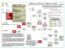
The information related to this issue determined as part of the incident investigation can be captured within a Cause Map, a visual form of root cause analysis. The first step of the process is to determine the impacts to the goals. In this case, both patient and employee safety are impacted due to the risk of illness from exposure to the particulates. The environmental goal is impacted because of the release of the particulates into the facility. Patient services are impacted because patients are being sent to other facilities as sterile procedures are not being performed (an impact to the production/ schedule goal). The labor and time required for an investigation is also an impact to the goal.

The second step of the process is the analysis: determining why these goals were impacted. The release of the particulates into the facility is because there are particulates within the air ducts, and the air ducts open into the facility to provide heating, ventilation and air conditioning. In order to determine where the particulates come from, first it must be determined what they are composed of. An environmental analysis determined that the particulates were rust, crumbling concrete, fiberglass fibers, and cladosporium (a common mold).

The analysis also identified that rust in air systems typically results from aged equipment exposed to moisture. Cladosporium also results from exposure to moisture. The air duct system pulls in outside air, including humidity, resulting in the system being exposed to moisture. The VA hospital is 45 years old, which actually makes it one of the “newer” VA facilities. (According to the VA, about 60% of its facilities are more than 60 years old.) While it’s unclear what maintenance or replacements have been performed on these components over the life of the facility, deferred maintenance is a general problem at VA facilities. According to the VA inspector general, there is a $10-12 billion maintenance backlog at the department.

Once the causes of the problems (or impacted goals) have been determined, the last step is to implement action items to reduce the risk of the problem recurring. There are two parts to this step: brainstorming possible solutions, and determining which will be most effective to meet the organization’s needs. The hospital considered bringing in mobile surgical units and installing high efficiency particulate air filters in the vents in the operating rooms. The cost of the mobile surgical units (over $70,000 per month) led the hospital to select only the solution of the air filters. At least one operating room is expected to be ready to return to service June 1st.

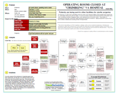
To view a one-page downloadable PDF of the incident investigation, including the impacted goals, analysis with evidence, and possible solutions, please click on “Download PDF” above.

After Patient Death, CMS Surveyor Declares “Immediate Jeopardy” To Patient Safety

By ThinkReliability Staff

From the moment a patient arrived at an endoscopy clinic in New York on August 28, 2014, things didn’t follow the usual procedure.  The patient brought her own ear, nose and throat physician (ENT) to accompany her into surgery for an esophagogastroduoudenoscopy (EGD), though the ENT did not have privileges at the facility.  The patient signed a consent form for anesthesia, and the EGD and her vital signs were taken, though it appears her weight was either not taken or not recorded.

After a time out that was initiated by the endoscopy technician (as opposed to the anesthesiologist, as required by policy) for the EGD, the patient was administered Propofol for sedation.  After the patient was sedated, the ENT attempted to perform a nasolaryngoscopy, despite not having facility privileges or a record of patient consent, but the initial attempt appears to have been unsuccessful.  After the EGD was completed, the ENT performed another nasolaryngoscopy beginning at 8:28 AM and ending (per interview records) at 8:30 AM.

By that time, the patient’s condition was quickly deteriorating.  Her blood pressure had dropped from its pre-procedure level of 118/80 to 84/40, her pulse from 62 to 47, and oxygen saturation from 100% to 92%.  Both the Cardiac Arrest Record and the Endoscopy Code Blue Record indicate that at 8:28 AM (the same time the second nasolaryngoscopy was beginning) the patient went into cardiac arrest/ ventricular tachycardia and measures were taken for resuscitation (including assisted ventilation, chest compressions, and administration of epinephrine and atropine).

The record of the surgery note that the laryngoscope was withdrawn at 8:30 AM, at which time cardiopulmonary resuscitation (CPR) was undertaken.  The patient was resuscitated and transferred to a hospital, where she died on September 4, 2014.  The cause of death from the autopsy report was anoxic encephalopathy (brain damage) caused by hypoxic cardiac arrest (oxygen deprivation).

The day prior to the patient’s death, a surveyor from the Centers for Medicare and Medicaid Services (CMS) declared “Immediate Jeopardy” due to “significant findings . . . which compromised patient safety”.  Specifically, the surveyor noted that the facility had risked patient safety by allowing a doctor without privileges to be allowed in the operating room and perform a procedure, and not obtaining consent or performing a time out for a procedure that was performed.

The facility quickly submitted a corrective action plan that revised procedures allowing visitors to the facility, ensuring informed consent and time out procedures are used before every procedure, and providing training on these updated procedures to staff.  Immediate jeopardy was removed on 9/5/14, although the facility was still considered out of compliance with CMS requirements, and was given until March 2 to maintain its certification.  (CMS has not released whether the facility has been successful.)

A lawsuit is underway that may provide more detail as to how the CMS findings caused (or didn’t cause) the patient’s death.  At this point, what is known can be captured in a timeline (for a chronology of events) and a Cause Map (to capture the cause-and-effect relationships that led to the impact to the goals) to start organizing and presenting information logically.  As more information is available, the Cause Map can be updated.

To see the timeline and initial Cause Map, click on “Download PDF” above.


65 surgeries required for boy who swallowed button battery

By ThinkReliability Staff

As you can imagine, as someone who regularly analyzes and reports on disasters of every kind, my mind is never far from things that could harm my children. Though the types of events I typically include are well-publicized, the real dangers for children tend to be things closer to home, that one might reasonably consider harmless.

Even after our blog about the dangers of children swallowing batteries (regarding a study that said a child in the US visits an emergency room every 3 hours for issues involving a battery and that 84% of these are button batteries), I didn’t really get it. Sure, swallowing anything is bad, and batteries have nasty chemicals in them. But it wasn’t until I read the story of Emmett Rauch that I really got how bad these issues could be.

When Emmett was one, he swallowed a button battery. His parents could tell that something wasn’t right, and Emmett was diagnosed with a cold, then croup. Luckily at a pediatrician visit 3 days after he swallowed the battery, the pediatrician had second thoughts and sent Emmett to the ER for an x-ray. Once the button battery was discovered lodged in his esophagus, the rush to treat him began. Emmett would receive 65 surgeries over the next four-and-a-half years to rebuild his esophagus and vocal chords.

Amazingly, Emmett is a survivor. He’s one of the lucky ones. There have been 15 deaths associated with small batteries over the last 6 years. Emmett’s mother, Karla Rauch, is now an activist for button battery ingestion awareness. The issues resulting in deaths and injuries to children (primarily under the age of 5) regarding button batteries are as follows:

Chemical reaction caused by batteries in the esophagus: Batteries (even “dead” ones) contain chemicals that create current. The moistness of the esophagus can cause a chemical reaction that can burn holes in the tissue.

Accessibility of button batteries: Kids like shiny things and they like to put things in their mouth. The first line of defense is preventing access to small batteries. Here’s how:

– Keep loose batteries out of reach of children

– Ensure battery compartments on products are secured

– Buy products with battery compartments that require a tool to open if possible

– Use duct tape to secure products with batteries that don’t require a tool

Difficulty of diagnosis – at home: Because the batteries are so small, kids will likely still be able to breathe after swallowing them, limiting parent’s ability to figure out that they’ve swallowed something they shouldn’t. Because of the ubiquity of small batteries, parents may not realize they’re missing.

Difficulty of diagnosis – at the hospital: An x-ray is required to determine that a child has swallowed a battery. An x-ray may not be called for if a doctor thinks (as is common) that the coughing or apparent throat damage is due to another sickness. Even though button batteries have been around for a while, they’re still not a risk that is very apparent to most people. So, if you think there is a possibility your child may have swallowed a battery, act quickly:

– Seek immediate medical attention if you believe a battery has been swallowed

– Do not let the child eat or drink

– Do not induce vomiting

– Tell the medical staff it may be a small battery

– If possible, provide information on the battery

To view an overview of this issue and solutions, please click on “Download PDF” above. Thanks to Karla Rauch for sharing her story. To learn more, see www.emmettsfight.com.

Deaths and Arrests Leave Many Questioning Mass Sterilizations

By ThinkReliability Staff

The deaths of 13 women after they underwent tubal ligation (female sterilization surgery) at a government- run clinic in India have resulted in outrage and arrests, but few answers. It is known that shortly after one of these mass sterilizations on November 8th at a mobile clinic, the women became ill and died.  The doctor was arrested for performing 83 sterilizations in 6 hours when government regulations permit only 30 in a day.  (The doctor says that pressure and incentives from the government encourage more sterilization surgeries despite guidelines.)  The cleanliness of the tools used for the surgeries as well as of the clinic itself are believed to have caused infections that led to severe septic shock that resulted in these deaths.

However, patients from different clinics who had taken antibiotics from the same batch used at the sterilization clinic also became ill, raising the question of contaminated – or outright counterfeit – drugs.  The owner and son of the manufacturing clinic that provided the antibiotics were also arrested.  It’s now believe that the women exhibited signs of potential poisoning and that trace amounts of a chemical used in rat poison were found in tests of the batch of antibiotics.  The signs of poisoning and septic shock can be similar, so additional testing is needed.

Either way, it’s clear that the care these women received was substandard.  Not only is the condition of the clinics and medication in question, but also the use of these surgeries as a form of family planning, which is common in the area.  In a survey, 34% of households indicated that female sterilization was their main method of family planning. Availability of less permanent birth control and medical care are limited, and population is booming.  The government has encouraged female sterilization, not only by running the mobile clinics but also paying the women who are sterilized.  The women who died from the procedure were given 1,400 rupees, or about $23 US.

Although there is currently limited information on what exactly lead to the death of these women, an analysis of those things that went wrong can still be useful to look for solutions.  (Click “Download PDF” above to view the one-page overview of a Cause Map, or visual root cause analysis, of what is known so far.)  Each possible cause (which indicates that more evidence is needed) can be developed with additional detail when more information is available.

Post-mortem inspection of the women who were killed indicated that their deaths were due to septic shock from severe infections.  The infections were believed to have been obtained during the surgery due to dirty tools or poor conditions.  (It’s not surprising that a clinic with limited resources known for having substandard cleanliness would struggle to ensure clean equipment for 83 surgeries in 6 hours.)  If the antibiotic they were given in order to fight potential infection was adulterated (and not providing the desired active ingredients, or too little of them to be effective), it would decrease the women’s ability to fight off the infection.

The potential poisoning (supported by sickness of others who received medication from the same batch) is believed to be due to counterfeit or adulterated medication.  Preliminary testing showed the presence of a chemical used in rat poison within the medication.  Counterfeit and adulterated drugs are both a problem in India; the World Health Organization (WHO) estimates that as many of 1 in 5 drugs made in India are fake.  The US FDA prevents the importation of the antibiotic used at the clinic from India.  Drug inspectors in the area indicate that about 25% are of substandard quality due to poor manufacturing practices.  Enforcement of drug quality is limited in the area, and issues with medication are seen frequently.

As mentioned above, both the surgeon and the owners of the pharmaceutical manufacturing facility have been arrested.  Both sterilizations and the use of certain drugs have been limited in the area until the causes resulting in the women’s deaths have been determined.  What hasn’t happened (yet) is a serious look at the country’s method of family planning in hopes that these types of tragedies never happen again.

Surgical Tool May Spread Cancer

By ThinkReliability Staff

Women who suffer from painful uterine fibroids may have their uterus removed for relief.  The most common hysterectomy procedure involves a 3-to-7-inch incision through which the entire uterus is removed intact.  Surgeries that can be performed laparoscopically through a smaller incision offer less scarring, fewer complications and shorter recovery time.  In order to remove the uterus through a smaller incision, it first has to be cut up into pieces.  This is performed using a laparoscopic power morcellator.  The device can also be used to remove fibroids only (called myomectomy) in a laparoscopic procedure.

On April 17, 2014, the Food and Drug Administration (FDA) issued a statement saying it “discourages the use of laparoscopic power morcellation during hysterectomy or myomectomy for uterine fibroids.” An advisory panel then provided a variety of recommendations, some going so far as to recommend completely banning the use of the device.

The issues surrounding the use of the morcellator can be examined in a Cause Map.  The Cause Map allows a visual diagram of cause-and-effect relationships that include the impacts associated with a particular issue.  In this case, the use of the power morcellator is found to increase the risk of spreading cancer, a clear impact to the patient safety goal.  Concerns about the delay in determining the safety risk (morcellators have been used since the 1990s) can be considered an impact to the patient services goal.  Sales of morcellators have been stopped, and as discussed above, the use of morcellators is being questioned by the FDA.

Beginning with the impacted goals, we can ask “Why” questions to determine the cause-and-effect relationships.  The risk for spreading cancer results from undetected cancer in tissues that are then cut up inside the body.  Uterine cancer is difficult to detect with available testing and can be difficult to distinguish from fibroids.  Additionally, cases have been found where cancer was missed in testing prior to surgical treatment.  Recent studies have shown that women who seek such surgeries have a higher incidence rate of uterine cancer (1 in 368 post-hysterectomy patients were found to have uterine cancer versus 1 in 10,000 for the general population).

The morcellator cuts up tissue so it can be removed laparoscopically in a minimally invasive hysterectomy or myomectomy for fibroids.  Until recently, there was no data or warning indicating that morcellators weren’t safe to use.  The device did not provide clinical trial data, which is not required for new products considered “substantially similar” to existing products that are already marketed.  The FDA does not believe that the cancer risk would have been discovered even with a higher level of scrutiny.  The FDA depends on a database of medical-device problems reported by doctors.  However, doctors are not required to report issues.  In addition, it’s not clear that many made the link from the spread of cancer after the use of the morcellator.  Or, some healthcare professionals say they didn’t report it because they believed the undetected cancer – not the use of a device – was to blame.

The link over spreading cancer has now clearly been established.  (Data is still being examined to see if the device worsens cancer as well.)  The FDA is working to determine what – if any – restrictions will be placed on morcellator use.  It’s pretty clear to some, especially given that this treatment for fibroids is considered elective and there are alternatives, including a traditional hysterectomy.  Says Dr. Craig Shriver, surgical oncologist at Walter Reed National Military Medical Center, “There is at present no safe way to offer laparoscopic power morcellation as part of any minimally invasive surgery.”

However, others think the tool still has a place for minimally invasive surgery and that patients should be able to determine for themselves if the risk is worth taking.  According to Dr. Jason D. Wright, the lead author of a study regarding morcellators published in the Journal of the American Medical Association study, “I don’t know that necessarily morcellation should be banned. But this data is important to allow people to make decisions.”

In the meantime, some facilities are attempting to enclose the uterus in a protective bag before morcellation to minimize the risk of tissue remaining in the body, but there is no evidence that this reduces the risk of cancer spread.  Until the FDA’s final decision, each patient and healthcare facility will have to do their own risk assessment to determine whether or not the tool should be used.


Alleged Radiology Misreading Results in Removal of Cancer Patient’s Healthy Kidney

By ThinkReliability Staff

On January 17, 2013, a radiologist discussed the results of a CT scan with an urologist.  The CT scans identified cancer in the kidney of an urologist’s patient.  Two months later, the patient underwent surgery to remove the kidney.  The kidney was examined by a pathologist, who declared it cancer-free.  The wrong kidney had been removed, allegedly due to a misidentification by the radiologist.

Wrong-site surgeries like this one can lead to severe patient safety consequences, as well as severe financial and regulatory consequences for the doctors and healthcare facilities involved.  This is why surgery performed on the wrong body part has been identified as a “never event“, or an event that should never occur in a healthcare facility.

Even with this designation and the known seriousness of the issues, wrong-site surgeries continue to occur.  The Joint Commission estimates that the prevalence of wrong-site surgeries in the United States is as high as 40 per week.

Clearly, action must be taken to reduce the risk of wrong-site surgeries.  To identify areas of potential improvement, it can help to look at an example of an actual case of wrong-site surgery to determine lessons learned.  We will examine the case of the wrong kidney being removed as an example of issues that can lead to wrong-site surgeries using the Cause Mapping method of root cause analysis.

It’s important to identify the impacts to the goals as a result of an incident.  In this case, the patient safety goal was clearly impacted as the patient now has only 3/4 of a kidney remaining, with the potential to cause serious health impacts.  (A portion of the cancerous kidney was removed in a later operation.)  The compliance goal is impacted because of the occurrence of a “never event” as discussed above.  The patient services goal is impacted due to the removal of the wrong (healthy) kidney.  The radiologist and urologist involved in the issue have been sued for more than $1 million as a result of the issue.  If all these issues received similar lawsuits, the costs to the health system would be over $2B a year.

Once the impacts to the goals are identified, asking “why” questions develops the cause-and-effect relationships that led to the issue.  In this case, the removal of the wrong kidney is alleged to have resulted from the radiologist misreading the CT scan that identified the kidney with cancer and passing that information on to the urologist who performed the surgery.  Clearly the urologist’s physical exam (if any) did not adequately determine the site of the cancer.

To better understand the steps that led to the surgery, they can be diagrammed in a Process Map.  A Process Map lays out a process in much the same way that a Cause Map visually lays out cause-and-effect relationships.  A very high level overview of the process used in this case is shown on the downloadable PDF.  What’s important to note is that an incorrect reading of a CT scan or other diagnostic tool propagates through the process.  With no second opinions or double checks built in, the diagnosis of cancer in the left kidney was the only information the urologist had to determine the operating site.

There are of course other errors in the surgical preparation procedure that can also cause wrong-site surgeries.  (Many of these errors are identified in our proactive write-up on wrong-site surgeries.)  As stated by Mark R. Chassin, M.D., President of The Joint Commission, “Wrong site surgery events occur basically because none of the processes that we use in taking care of patients is perfect.”  Equally important is that the people performing the processes are not perfect.  Although both processes and people’s performance can be improved, it will never reach perfection.  For this reason, adding double checks and second opinions into processes is essential to reduce the risk of the one mistake resulting in a devastating patient safety impact.  In this case, having a second opinion on the CT scan, or having the physician re-identify the area with a physical exam prior to surgery (if possible) may have identified the error prior to removal of a healthy kidney.

View the Cause Map and process map by clicking on “Download PDF” above.

13-Inch Surgical Tool Left In Patient for Months

By Kim Smiley

For about two months after surgery to remove a large malignant tumor Donald Church complained of severe pain.  Initially, he was told that it was normal pain associated with recovery from a major surgery, but an x-ray was ordered after his physician felt a lump in his abdomen.  The x-ray revealed that a malleable retractor similar in size to a ruler had been left inside his body after surgery.  A second surgery was done to remove the tool.  Mr. Church is not expected to suffer long-term health consequences and received a $97,000 settlement.

A Cause Map, a visual format for performing a root cause analysis, can be built to help understand how this issue happened.  Once all the causes that contributed to an issue are found, potential solutions can be found and the most promising can be implemented to help reduce the risk of a similar issue reccurring.

So how did this happen?  How does a large surgical tool get left inside a patient?  This occurred because the patient needed surgery to remove a tumor, the malleable retractor was used during the surgery and the surgeons were unaware that the tool remained inside the patient. (These causes are vertical on the Cause Map with “and” between them because all 3 were necessary for the issue to happen.)  A malleable retractor was used while the wound was being closed to help protect the organs under the wound from possible puncture from the suturing needle.

The surgeon was unaware that the tool was inside the patient because he couldn’t see it and there wasn’t an adequate system in place to manage surgical tools.  Malleable retractors are normally held partly out of the wound, but it had slipped entirely inside the wound during the surgery.  Once the tool was out of sight, it was forgotten.  While many hospitals have requirements to formally count surgical tools as they enter and leave the operating room to ensure that all are accounted for, there wasn’t a policy in place in the facility that performed the surgery at the time.  With no formal system to track tools, there weren’t any easy indications to the operating team that there was a problem.

While this is a particularly egregious example, there are an estimated 4,000 cases of retained surgical items each year in the United States. Better solutions need to be found to reduce the risk of this preventable and potentially deadly problem from happening.  A simple solution to reduce the risk for retained surgical items is to institute a formal procedure for counting surgical supplies and tools before and after surgery.  Simple manual counts are a first step, but errors still occur, especially in the often hectic and stressful environment in an operating room.  Some hospitals use a visual inventory system where tools are brought in a special storage bag with an individual compartment for each item.  As items are done being used they are put back into their specific spot.  If all compartments are full, everything is accounted for so it’s easy to tell if something is missing.

Another solution that is gaining in popularity is use of an electronic tracking system.  The most common use of electronic systems is to track sponges, which are by far the most common object left inside patients.  Each sponge has an electronic tag and the patient is scanned after surgery to verify that none were left behind.  Sponge tracking systems add about $8 to $12 to the cost of each surgery and have dramatically reduced the number of retained sponges when used.

To view a high level Cause Map, click on “Download PDF” above.

The Willie King Case: Wrong Foot Amputated

By Kim Smiley

In one of the most notorious medical error examples in US history, the wrong foot was amputated on a patient named Willie King on February 20, 1995.  Both the hospital and surgeon involved paid hefty fines and the media had a feeding frenzy covering the dramatic and alarming mistake.

So how did a doctor remove the wrong foot?  Such a mistake seems difficult to comprehend, but was it really as mind boggling as it looks at first glance?

The bottom line is that the doctor honestly believed he was removing the correct foot when he began the surgery. The blackboard in the operating room and the operating room schedule all listed the wrong foot because the scheduler had accidentally listed the wrong foot.  After reading the incorrect paperwork, the nurse prepped the wrong foot.  When the doctor entered the operating room, the wrong foot was prepped and the most obvious documentation listed the wrong foot.  Basically, the stage was set for a medical error to occur.

The foot itself also looked the part.  The patient was suffering from complications of diabetes and both of his feet were in bad shape.  The “good” foot that was incorrectly removed looked like a candidate for amputation so there were no obvious visual clues it wasn’t the intended surgery site. Other doctors had testified in defense of the doctor saying the majority of other surgeons would have made the same mistake given the same set of circumstances.

There was some paperwork that listed the correct foot to be amputated, such as patient’s consent form and medical history.  This paperwork was available in the operating room, but no procedures in place at the time required the doctor to check these forms and these forms were far less visual than the documents where the incorrect information was listed.  Additionally, the doctor never spoke directly with the patient prior to the surgery which was another missed opportunity for the mistake to be caught.

Clearly the procedures needed to be changed to prevent future wrong site surgeries from occurring and a number of changes have been incorporated in the time since this case occurred to help reduce the risk of this type of medical error.  Surgeons in Florida are now required to take a timeout prior to beginning a surgery.  During the time out they are required to confirm that they have the right patient, right procedure and right surgical site.  This rule has been in place since 2004.

Mistakes will always happen, such as numbers being transposed or misheard words over the phone, but small mistakes need to be caught before they become big problems. Procedures like a timeout can significantly reduce the likelihood of an error going uncorrected.  In an ideal world, the simple mistake by the scheduler would have been caught long before it culminated in a surgery on the wrong body part.

A visual root cause analysis, called a Cause Map, can be built to illustrate the facts of this case.  A Cause Map intuitively lays out the cause-and-effect relationships including all the causes that contributed to an issue.  To view a Cause Map of this example, click on “Download PDF” above.

Patient Receives Unneeded Mastectomy

By ThinkReliability Staff

Information about a lab mix-up that resulted in a patient receiving an unnecessary mastectomy was recently released by a health authority in Nova Scotia, Canada.  The authority has conducted an investigation into the error and how it occurred.  We can look at some of the information that will need to be considered in order to reduce the risk of a similar issue recurring.

First, we capture the “What”, “When”, and “Where” of the incident.  In this case, a switch of pathology results occurred in late April, 2013 at a cancer center in Nova Scotia.  The error was caught as a result of oversight analysis of tissue samples.

We can capture the goals impacted as the result of the issue.  In this case, the patient safety goal was impacted because a patient (Patient 1) received an unnecessary surgery (mastectomy).  In addition, the patient safety goal was impacted because another patient (Patient 2) did not receive a necessary surgery.  The staff employees are impacted because they are reportedly devastated, as frequently happens in cases like these.  The organization goal is impacted due to the apology given to the public as a result of this issue.  The patient services goal is impacted due to a switch of the tissue samples.  The property goal is impacted because an unnecessary procedure was performed, and the labor goal is impacted due to the extensive investigation that is taking place.

Asking “Why” questions can help determine the cause-and-effect relationships that led to the impacted goals.  In this case, the patient safety impacts are due to the switching of the patient’s sampling.  This occurred due to the results being recorded into the wrong records.

While trying to solve a problem, it can be helpful to examine the related processes.  In this case, we look at the tissue sampling process.  Any process is meant to get from point A to point B. In this case, the process ideally takes us from a tissue sample being taken (point A) to a diagnosis (point B).  We know that we did not get to point B in this case (i.e. the diagnosis was incorrect).  Looking at the steps in more detail can help us determine which specific part of the process did not go as intended, which will allow us to identify process-specific solutions.

A sample is taken from a patient, labeled, and sent to the lab.  The lab tests the sample, obtains the results, then delivers them to the patient’s physician or care center.  At that point, the results are recorded in the patient’s records and then used to make a diagnosis.  The error reportedly occurred at the point where the results were entered into the patients’ records.

Once we’ve identified the specific point where the error occurred, we can identify potential solutions.  In this case, the facility involved is implementing bar-coding and moving towards an automated system.  Although there is still the potential for error, it is reduced with automated systems and bar codes because the data has to be transcribed fewer times. As the Premier of Nova Scotia stated, “Human error is always a possibility. But one of the things we strive for is to ensure there are appropriate controls in place to ensure that the risk of these things is absolutely minimized.”

To view the Cause Map and Process Map, please click “Download PDF”.

A Tongue Tie Release Wrongly Performed in Case of Tongue Lesion Resection

By ThinkReliability Staff

A California hospital has been fined $50,000 – its fifth administrative penalty from the State since 2009 – for performing the wrong procedure on a 6-year-old boy.  The boy was supposed to receive a tongue lesion resection, but instead a tongue tie release was performed.

We can examine the issues that resulted in this incident within a Cause Map, or visual root cause analysis.  The first step in any analysis is to define what you are analyzing.  We begin with impacts to the organization’s goals.  In this case, we look at the impacted goals from the respect of the hospital.  First, the patient safety goal  was impacted due to an increased risk of bleeding, infection, and complications from anesthesia.  The compliance goal is impacted because performing the wrong surgical procedure on a patient is a “Never Event” (events that should never happen).  The organizational goal is impacted because of the $50,000 fine levied by the State of California.  The patient services goal is impacted because the wrong procedure was performed and the labor goal was impacted due to the additional procedure that was required to be performed.

The second step of our analysis is to develop the cause-and-effect relationships that describe how the incident occurred.  We can develop these relationships by beginning with the Impacted Goals and asking “why” questions.  For example, the patient safety goal was impacted because of the additional risk to the patient. The patient received additional risk because of the performance of an additional procedure.  An additional procedure was necessary because the wrong procedure was initially performed.

There are many causes that contributed to the wrong surgery being performed.  These causes are outlined in the  report provided by the California Department of Public Health.  In this case, there were several causes that likely resulted in the wrong procedure.  The Operative Report had the incorrect diagnosis – tongue tie – which would suggest that a release would be the appropriate procedure.  Additionally, the Anesthesia Record contained the wrong procedure (tongue tie release), possibly because the Pre-Anesthesia Evaluation originally noted that a tongue tie release was to be performed and was later corrected (by crossing out the incorrect procedure and writing in the actual procedure).

The type and site of surgery was not verified.  The surgeon who performed the surgery could not remember if a time-out had been performed, although there was a record of a time-out performed immediately prior to the surgical procedure.  Since the time-out was performed immediately prior to the procedure and the surgeon was unable to remember the proper procedure, the time-out was obviously ineffective.

The surgeon stated after the surgery that he believed that the tongue tie release surgery which was performed was indicated based on scar tissue that was found under the tongue.  The surgeon did not notice the lesion on the tongue during the surgery and no pre-surgical exam was performed by the surgeon.  Additionally, the surgical site was not marked (as the site of the correct, as well as the incorrect, surgeries were both within the patient’s mouth).

During the procedure, none of the other staff stopped the surgery as it was occurring.  However, given the proximity of the “correct” site to the “incorrect” site, it may have been difficult for the other staff to see what was going on.  The surgeon did note that the lesion removal should have created a sample, the lack of which was not noted by staff.

The surgeon involved in this case has indicated that he will be examining his patients prior to surgery in the future. Hopefully this incident will also serve as a reminder to all medical staff that in the case of a site that cannot be marked as per procedure, extra care should be taken to ensure the correct site is operated on and the correct procedure is performed.

To view the Outline and Cause Map, please click “Download PDF” above.  Or click here to read more